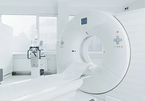
Computed Tomography is a diagnostic imaging technique that examines every part of the body with extremely detailed images. It is an X-ray examination that collects data through the passage of X-ray beams in the selected area. These are processed by a computer in order to render a three-dimensional image.
Ticino Animal Hospital with this service, thus becomes one of the first centers in Switzerland .
How are CT Scans Done?
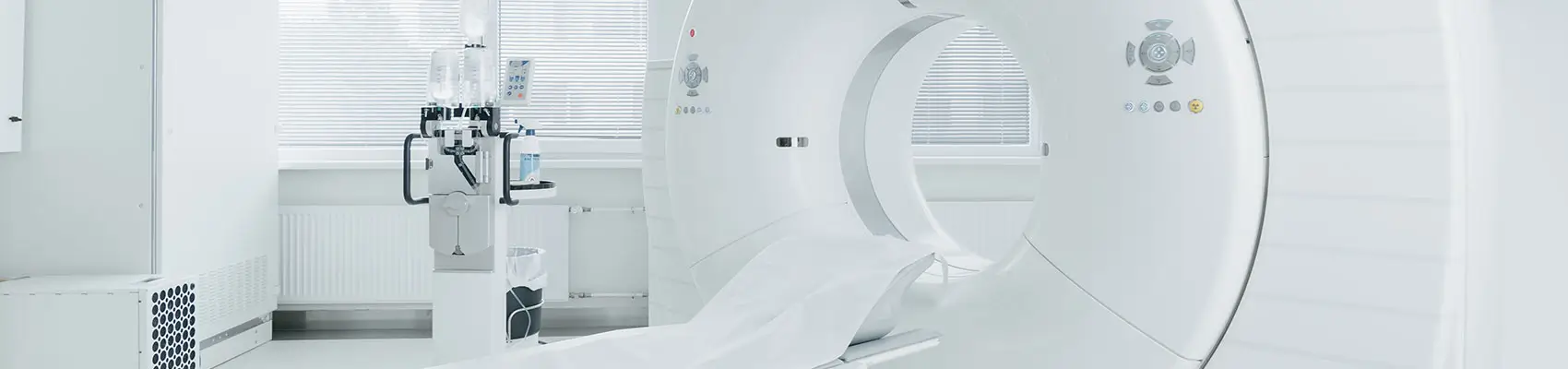
The X-ray tube, which produces X-rays, rotates around the patient. The patient lies on a table that moves horizontally and passes through a circular machine. Sometimes, to get better images of the vascular system (arterial and venous) of organs and tissues, an iodine-based contrast medium is given intravenously.
What are some of the most common indications?
In Oncology for tumor staging, CT-guided biopsies, and planning complex surgical procedures. In polytraumatized patients, for example following car accidents or falls from height, to assess skeletal conditions, the brain, and thoracic and abdominal organs. In Orthopedics for studying various forms of hip, shoulder, and elbow dysplasia, complex fractures, and facial trauma. In Neurology for evaluating intracranial and spinal conditions (spinal trauma, herniated discs, inflammatory diseases). To increase diagnostic sensitivity for thoracic and abdominal conditions that are difficult to detect with other diagnostic tools, such as lobar torsion, ectopic ureter, and portosystemic shunt. Evaluation of the nasal cavities and frontal sinuses, the retrobulbar space, and the middle and inner ear.
What are the contraindications in dogs and cats?
Since CT uses X-rays, the examination is contraindicated during pregnancy and in patients allergic to contrast agents.
The use of contrast agents must be carefully evaluated in patients with diseases affecting organ function such as the kidneys or liver (diabetes, renal failure, hepatic failure).
How should you prepare for this exam?
In veterinary medicine, CT scans are generally performed under general anesthesia, except in specific cases. This is necessary not only to immobilize the patient but also to obtain optimal diagnostic images. For this reason, and to allow the administration of contrast agents, the patient must have undergone complete blood tests.
Additionally, it is important that the patient fasts for at least 8 hours (water and regular medications are allowed before the exam, as they do not interfere with the procedure).
How is the exam performed and how long does it take?
A few days before the scheduled exam, a consultation with the radiologist will take place to understand the exact reason for the exam and optimize the imaging protocol. All clinical and diagnostic documentation will be collected (medical records, imaging exams, specialist reports) along with the prescription from your referring veterinarian.
It is important to report any history of allergic reactions (seasonal, food-related, or drug-related).
All exams are performed as day hospital procedures to allow post-anesthesia monitoring and ensure patient safety.
The duration of the exam varies depending on the anatomical region and diagnostic suspicion and generally takes between 10 and 20 minutes. During the entire procedure, your pet’s vital functions will be continuously monitored by a dedicated anesthesia team.
After the exam, unless complications arise, the patient is discharged, can return home, and resume normal activities immediately. Food and water intake will follow the anesthesiologist’s instructions.
How to book?
Appointments are arranged directly with the responsible radiologist by calling +41 (0)91 225 3765. When booking, it is advisable to have the referral from your veterinarian available.
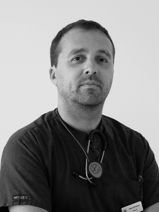
Dr. Paolo Martegani (Second-level Master’s degree in Ultrasonology of dogs and cats) is the head of the department and coordinates diagnostic activities.